Clicky

Ok

En poursuivant votre navigation sur ce site, vous acceptez l'utilisation de cookies. Ces derniers assurent le bon fonctionnement de nos services. En savoir plus.

10/11/2012

Exsufflation à l'aiguille: Pas si efficace ?

Does needle thoracostomy provide adequate and effective decompression of tension pneumothorax ?

Martin M et All.J Trauma Acute Care Surg. 2012;00: 00Y00 (DOI: 10.1097/TA.0b013e31825ac511) 

Il existe un grand débat sur les modalités d'exsufflation d'un pneumothorax suffocant (site de ponction, longueur et calibre de l'aiguille, thoracostomie au doigt, drainage ?).

Ce travail expérimental pointe du doigt, UNE FOIS DE PLUS, les limites de l'exsuffflation à l'aguille:

"All NTs were patent on initial placement, but 5 (26%) demonstrated mechanical failure (due to kinking, obstruction, or dislodgment) within 5 minutes of placement, all associated with hemodynamic decline. Among the 14 NTs that remained patent at 5 minutes, 6 (43%) failed to relieve tension physiology for an overall failure rate of 58%"

PTX REleif.JPG

Ces données doivent pousser plus avant la formation vers les deux autres moyens que sont l'exsufflation au doigt (présenté par beaucoup comme la méthode de référence) et bien sûr le drainage thoracique.

Approfondir: Document 1  Document 2  La réflexion US TCCC

01/10/2012

Epaisseur de paroi thoracique: Le point chez le militaire français

Mesure de l’épaisseur de la paroi thoracique chez des militaires français :

Quelle technique pour l’exsufflation du pneumothorax compressif lors du sauvetage au combat?

A propos d’une étude descriptive réalisée du 1er Mars au 15 Avril 2010

au 7ème Bataillon de Chasseurs Alpins de Bourg Saint Maurice.

LAMBLIN A. Thèse Lyon 2012


Le diaporama de présentation      :       EpThoDiaporama.pdf

La thèse                                          :       Lien

20/09/2012

Pneumothorax: La sonde d'écho à quel endroit ?

 Anatomical distribution of traumatic pneumothoraces on chest computed tomography: implicationsfor ultrasound screening in the ED

Mennicke M et alll. Am J Emerg Med 2012 Sep;30(7):1025-31

Un travail qui précise les zones optimales d'exploration de la région thoracique pour l'identiifcation optimale des pneumothorax traumatiques.

----------------------------------------------------------------------------------------

OBJECTIVES:

We sought to assess the anatomical distribution of traumatic pneumothoraces (PTXs) on chest computed tomography (CT) to develop an optimized protocol for PTX screening with ultrasound in the emergency department (ED).

METHODS:

We performed a retrospective review of all chest CTs performed in one ED between January 2005 and December 2008 according to presence, location, and size of PTX. Pneumothoraces were then measured and categorized into 14 anatomical regions for each hemithorax.


RESULTS:

A total of 277 (3.8%) PTXs were identified, with 26 bilateral PTX, on 3636 chest CTs performed during the study period. Etiology was blunt (85%) or penetrating trauma (15%). Eighty-three (45%) PTXs were radiographically occult on initial chest x-ray. One hundred eighty-three (66%) PTX had no chest tube at the time of CT. For both hemithoraces, the distribution demonstrated increasing PTX frequency and size from lateral to medial and from superior to inferior. Region 12 (parasternal, intercostal spaces [ICS] 7-8) was involved in 68% of PTX on either side; region 9 (parasternal, ICS 5-6), in 67% on the left and in 52% on the right; and region 11 (lateral to midclavicular line, ICS 7-8), in 46% on the left and in 53% on the right. The largest anterior-to-posterior PTX dimension was seen in region 12.

zones thorax.jpg

Pneumothorax.jpg

CONCLUSIONS:

Our results indicate that 80.4% of right- and 83.7% of left-sided traumatic PTXs would be identified by scanning regions 9, 11, and 12. These findings suggest that a standardized protocol for PTX screening with ultrasound should include these regions.

----------------------------------------------------------------------------------------


15/05/2012

Paroi thoracique: En moyenne 3,06 cm chez le japonais

JapanseseChestWall 1.JPG

The mean CWT measured in 192 males and 64 females was 3.06  1.02 cm. The CWT values at 483 sites (94.3%) were less than 5.0 cm. The CWT of females was significantly greater than that of males (3.66 cm vs. 2.85 cm, p < 0.0001), and patients with subcutaneous emphysema had greater CWTs than those without it (4.16 cm vs. 3.01 cm, p < 0.0001).


JapaneseChestWall.JPG

16/04/2012

3 Côtés: La valve de Russel

Outre qu'elle présente un dispositif type pansement 3 côtés, cette vidéo est intéressante par la qalité de la présenttaion clinique d'une plaie soufflante.

le thorax se soulève

la fréquence est rapide

la respiration n'est pas harmonieuse

car l'ampliation est faible avec tirage intercostal sans ballotement abdominal

le thorax est ouvert

le thorax souffle

http://www.prometheusmedical.co.uk/uploads/videos/RCS-sim...


Une vidéo de présentation technique

11/03/2012

Drainage Thoracique: Histoire de drain

A portable thoracic closed drainage instrument for hemopneumothorax.J Trauma. 2012;72: 671–675 Tang H; Pan T MD et all.

La pose d'un drain thoracique en préhospitalier s'impose dès lors qu'un pneumothorax n'est pas traité de manière correcte par une thoracostomie au doigt/Exsufflation à l'aiguille ou si l'évacuation du blessé est retardée. Le Front Line Chest Tube est l'équipement préconisé par la procédure du sauvetage au combat. Une alternative de fortune peut être une sonde d'intubation ou une canule de trachétomie. Cet article illustre le travail de réflexion fait pour mettre à disposition un équipement adapté au contexte pré-hopsitalier notamment militaire.  L'emploi de tubes à ballonet est proposé par des auteurs chinois qui ont dévelopé ce nouveau matériel présenté dans le Journal of Trauma

NewChestDrainage.jpg

23/02/2012

Exsufflation à l'aiguille: Le risque est rare mais réel !

Subclavian artery laceration: A serious complication of needle decompression

Riwoe D et all. Emergency Medicine Australasia (2011) 23, 651–653

Un article qui rapporte la survenue d'une dialcération de l'artère sous clavière après exsufflation à l'aiguille par voie antérieure. Les auteurs militent pour la réalisation d'une thoracostomie au doigt par voie latérale ou après repérage échographique sinon l'emploi de repère cutanés préconisés par Wax et coll. qui font appel à la reconnaissance de la fourchette sternale et la ligne médio-thoracique mamelonnaire.

Epaisseur de paroi thoracique: Une de plus qui dit le contraire!

Anterior Versus Lateral Needle Decompression of Tension Pneumothorax: Comparison by Computed Tomography Chest Wall Measurement.

Sanchez LD et all.  Academic Emergency Medicine 2011; 18:1022–1026

Un article qui présente des données qui contredisent le caractère plus épais de la paroi thoracique antérieure versus la paroi latérale, du moins au niveau des repères classiques d'exsufflation/Thoracostomie. La discussion n'est donc pas close. L'intérêt de ce document est aussi d'alerter sur le fait que le recours à de long cathéter peut améliroier le taux de succès d'une exsufflation mais aussi le risque de ponction de structures vasculaires sous jacentes.

paroitho.JPG

31/01/2012

Le triangle de sécurité pour la thoracostomie

Chest drain insertion is not a harmless procedure – are we doing it safely ?

Elsayed H. et All. Interactive CardioVascular and Thoracic Surgery 11 (2010) 745–749

Près de la moitié des drains thoraciques ne sont pas posés au bon endroit

thorax,drainage thorax

thorax,drainage thorax 

On rappelle la notion de triangle de sécurité (Le BTS Guideline)

TriangleSecurité.JPG

http://www.surgeryjournal.co.uk/article/S0263-9319%2811%2...

La réalisation d'une thoracostomie au doigt et l'insertion d'un drain doit se faire de manière sécuritaire.

Le choix de la voie axillaire est certainement à privilégier.

17/01/2012

Drainage de la plèvre: Techniques et pièges

Lisez donc ce document. Une synthèse du drainage pleural à l'hôpital. Révisez les repères

http://www.srlf.org/rc/org/srlf/htm/Article/2011/4ce9bc02...


06/01/2012

Passer par le côté, c'est mieux

J Trauma. 2011 Nov;71(5):1099-103; discussion 1103.

Optimal positioning for emergent needle thoracostomy: a cadaver-based study.

Source

Division of Trauma and Surgical Critical Care, University of Southern California, Los Angeles, California 90033-4525, USA. kinaba@surgery.usc.edu

Abstract

BACKGROUND:

Needle thoracostomy is an emergent procedure designed to relieve tension pneumothorax. High failure rates because of the needle not penetrating into the thoracic cavity have been reported. Advanced Trauma Life Support guidelines recommend placement in the second intercostal space, midclavicular line using a 5-cm needle. The purpose of this study was to evaluate placement in the fifth intercostal space, midaxillary line, where tube thoracostomy is routinely performed. We hypothesized that this would result in a higher successful placement rate.

METHODS:

Twenty randomly selected unpreserved adult cadavers were evaluated. A standard 14-gauge 5-cm needle was placed in both the fifth intercostal space at the midaxillary line and the traditional second intercostal space at the midclavicular line in both the right and left chest walls. The needles were secured and thoracotomy was then performed to assess penetration into the pleural cavity. The right and left sides were analyzed separately acting as their own controls for a total of 80 needles inserted into 20 cadavers. The thickness of the chest wall at the site of penetration was then measured for each entry position.

RESULTS:

A total of 14 male and 6 female cadavers were studied. Overall, 100% (40 of 40) of needles placed in the fifth intercostal space and 57.5% (23 of 40) of the needles placed in the second intercostal space entered the chest cavity (p < 0.001); right chest: 100% versus 60.0% (p = 0.003) and left chest: 100% versus 55.0% (p = 0.001). Overall, the thickness of the chest wall was 3.5 cm ± 0.9 cm at the fifth intercostal space and 4.5 cm ± 1.1 cm at the second intercostal space (p < 0.001). Both right and left chest wall thicknesses were similar (right, 3.6 cm ± 1.0 cm vs. 4.5 cm ± 1.1 cm, p = 0.007; left, 3.5 ± 0.9 cm vs. 4.4 cm ± 1.1 cm, p = 0.008).

CONCLUSIONS:

In a cadaveric model, needle thoracostomy was successfully placed in 100% of attempts at the fifth intercostal space but in only 58% at the traditional second intercostal position. On average, the chest wall was 1 cm thinner at this position and may improve successful needle placement. Live patient validation of these results is warranted.

26/11/2011

Exsufflation: 5 cm suffisent VRAIMENT !

Determination of the appropriate catheter length for needle thoracostomy by using computed tomography scans of trauma patients in Japan
Injury, In Press, Corrected Proof, Available online 24 December 2010
Takeshi Yamagiwa, Seiji Morita, Rie Yamamoto, Tomoko Seki, Katsuhiko Sugimoto, Sadaki Inokuchi
PDF (164 K) 

Background

Previous studies reported a high failure rate in relieving tension pneumothorax by needle thoracostomy, because the catheter was not sufficiently long to access the pleural space. The Advanced Trauma Life Support guideline recommends needle thoracostomy at the second intercostal space in the middle clavicular line using a 5.0-cm catheter, whereas the corresponding guideline in Japan does not mention a catheter length. It is necessary to measure the chest wall thickness (CWT) and determine the appropriate catheter length taking the differences of habitus in race and region into consideration. This study was designed to analyse CWT in Japanese trauma patients by computed tomography and to determine the percentage of patients whose pleural space would be accessible using a 5.0-cm catheter.

Patients and methods

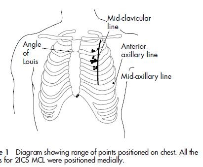
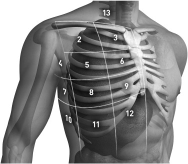
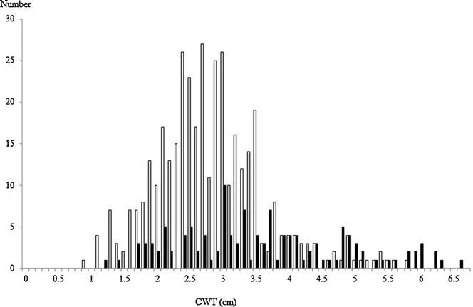
We performed a retrospective review of chest computed tomography of 256 adult Japanese trauma patients who were admitted to the level 1 trauma centre of Tokai University Hospital in Kanagawa, Japan between January and July 2008. In 256 patients, the CWT at 512 sites (left and right sides) was measured by chest computed tomography at the second intercostal space in the middle clavicular line. The frequency of measurement sites <5.0 cm was calculated simultaneously.

The samples were divided according to gender, side (left and right), abbreviated injury scale (<3, ≧3), arm position during examination (up/down), and the existence or non-existence of associated injuries (pneumothorax, subcutaneous emphysema, and fracture of the sternum and ribs); the CWT of each group was compared.

Results

The mean CWT measured in 192 males and 64 females was 3.06 ± 1.02 cm. The CWT values at 483 sites (94.3%) were less than 5.0 cm. The CWT of females was significantly greater than that of males (3.66 cm vs. 2.85 cm, p < 0.0001), and patients with subcutaneous emphysema had greater CWTs than those without it (4.16 cm vs. 3.01 cm, p < 0.0001).

Conclusion

The mean CWT at the second intercostal space in the middle clavicular line was 3.06 cm. It is likely that over 94% of Japanese trauma patients could be treated with a 5.0-cm catheter.

Exsufflation:Au bon endroit !

Réaliser l'exsufflation au bon endroit n'est pas si simple, même quand on est censé être un professionnel !

RepèreExsufflation.JPG

The right place in the right space? Awareness of site for needle thoracocentesis
E P Ferrie EP et all.
Emerg Med J 2005;22:788–789. doi: 10.1136/emj.2004.015107

La procédure

15/11/2011

Exsuflation par voie antérieure: Au 5ème axillaire plutôt qu'au 2ème antérieur?

Optimal Positioning for Emergent Needle Thoracostomy: A Cadaver-Based Study

Inaba K et all. J Trauma. 2011;71: 1099–1103

Un travail de plus sur la pertinence de la remise en cause de la recommandation d'aborder la paroi thoracique au 2ème espace intercostal sur la ligne médioclaviculaire. Ces repères sont en rapport avec une plus grande profondeur de l'espace pleural source d'échec de décompression pleurale lors d'exsufflation à l'aiguille. Certains proposent de réaliser cette dernière par voie latérale.  Un  travail de plus qui conforte cette approche.

-----------------------

Background: Needle thoracostomy is an emergent procedure designed to relieve tension pneumothorax. High failure rates because of the needle not penetrating into the thoracic cavity have been reported. Advanced Trauma Life Support guidelines recommend placement in the second intercostal space, midclavicular line using a 5-cm needle. The purpose of this study was to evaluate placement in the fifth intercostal space, midaxillary line, where tube thoracostomy is routinely performed. We hypothesized that this would result in a higher successful placement rate.

 Methods: Twenty randomly selected unpreserved adult cadavers were evaluated. A standard 14-gauge 5-cm needle was placed in both the fifth intercostal space at the midaxillary line and the traditional second intercostal space at the midclavicular line in both the right and left chest walls. The needles were secured and thoracotomy was then performed to assess penetration into the pleural cavity. The right and left sides were analyzed separately acting as their own controls for a total of 80 needles inserted into 20 cadavers. The thickness of the chest wall at the site of penetration was then measured for each entry position.

 Results: A total of 14 male and 6 female cadavers were studied. Overall, 100% (40 of 40) of needles placed in the fifth intercostal space and 57.5% (23 of 40) of the needles placed in the second intercostal space entered the chest cavity ( 0.001); right chest: 100% versus 60.0% ( 0.003) and left chest: 100% versus 55.0% ( 0.001). Overall, the thickness of the chest wall was 3.5 cm  0.9 cm at the fifth intercostal space and 4.5 cm 1.1 cm at the second intercostal space ( 0.001). Both right and left chest wall thicknesses were similar (right, 3.6 cm 1.0 cm vs. 4.5 cm  1.1 cm, 0.007; left, 3.5  0.9 cm vs. 4.4 cm  1.1 cm, p 0.008).

Conclusion: In a cadaveric model, needle thoracostomy was successfully placed in 100% of attempts at the fifth intercostal space but in only 58% at the traditional second intercostal position. On average, the chest wall was 1 cm thinner at this position and may improve successful needle placement. Live patient validation of these results is warranted.

Epaisseur.JPG

06/10/2011

Paroi thoracique: Epaisseur chez le français ?

Ce document rapporte la mesure de l'épaisseur de la paroi thoracique chez le militaire français, du moins dans la brigade alpine. Elle est en moyenne de 4,2 cm sur la ligne médio-claviculaire et de 3.03 cm en axillaire. 25% des combattants alpins ont une épaisseur > 5cm sur la ligne médio-claviculaire pour seulement 5.7% en axillaire. La conséquence en est la recommandation de réaliser les exsufflations en médecine de guerre non pas avec une aiguille de longeur supérieure à 5 cm mais par voie latérale. En effet le recours à la voie médio-claviculaire avec des aiguilles de 8 cm est à haut risque non seulement de ponction parenchymateuse mais aussi de strutures médiastinales vasculaires. Par ailleurs la réalisation d'une thoracostomie à la pince et au doigt doit être considérée comme le geste de référence, bien moins dangereux bien que de réalisation moins aisée.

Epaisseur Thorax.pdf

11/09/2011

Drainage thoracique: Prudence

Menger T et all.  doi:10.1016/j.injury.2011.06.420

La pose d'un drain thoracique n'est pas si simple que cela. C'est ce que rappelle cet article qui retrouve 28% de complications en cas de traumatisme ouvert et 17% en cas de traumatisme fermé.


Drain tHO.jpg

Donc pas de précipitation. L'urgence thoracique est à lever la compression par une thoracostomie au doigt. Un drain si l'EVASAN tarde.

26/12/2010

Drainage thoracique en images

Le drainage thoracique en condition de combat doit rester l'exception. Il peut se justifier en cas d'échec ou d'insuffisance de l'exsufflation ou de la thoracostomie au doigt. Il peut également s'envisager selon les conditions tactiques si le délai d'EVASAN es long. Le document ci-après présente de manière très didactique la réalisation de ce geste pas si anodin qu'il n'y paraît.

http://www.copacamu.org/IMG/pdf/3-roch.pdf


23/12/2010

Exsufflation: 5 cm suffisent !

Can J Surg. 2010 June; 53(3): 184–188

Thoracic needle decompression for tension pneumothorax: clinical correlation with catheter length

Ball CG et all.

"

Résumé

Needle thoracostomy decompression was attempted in 1.5% (142/9689) of patients. Among patients with blunt injuries, the incidence was 1.4% (101/7073). Patients transported by helicopter (74%) received a 4.5-cm sheath. The remainder (26% ground transport) received a 3.2-cm catheter. A minority in each group (helicopter 15%, ground 28%) underwent immediate chest tube insertion (before thoracic ultrasound) because of ongoing hemodynamic instability. Failure to decompress the pleural space by NT was observed via ultrasound and/or CT in 65% (17/26) of attempts with a 3.2-cm catheter, compared with only 4% (3/75) of attempts with a 4.5-cm catheter (p < 0.001).

" 

Morceau choisi

Although some authors13,16 have called for 7- to 8-cm needles to ensure that all OPTXs are decompressed, it appears that even catheters as short as 4.5 cm can puncture the heart at standard insertion locations in 2.5% of trauma patients.24 Other complications include chest wall hematoma, hemothorax, empyema and dislodgement in up to 8% of patients.18,25 In an attempt to avoid these issues, as well as access the pleural space more reliably, support for axillary NT is increasing.3,18,22,25,39 This lateral location takes advantage of a thinner chest wall (mean 2.6 cm)16 and is the military’s first choice if under fire because it allows medics to keep a soldier’s body armor in place while achieving decompression.40 Although we observed no direct complications in our study, we support the use of a catheter at of least 4.5 cm in length.

Commentaires

Le pneumothorax suffocant est la seconde cause de mort évitable au combat. Sa pris en charge repose sur la décompression de l'air retneu dans la cage thoracique. Outre la thoracostomie au doigt, geste de référence simple et sans danger, il peut être effectué une décompression à l'aiguille. Certains militent pour l'emploi d'un cathéter de grande taille de 8 cm du fait de l'épaisseur de la paroi thoracique, en particulier chez les militaires . Cependant le risque de plaie cardiaque et des gros vaisseaux est relativement important et peut atteindre 2.5% des blessés (1, 2, 3), ce d'autant plus que les ponctions sont habituellement réalisées de manière beaucoup trop médianes.

TensionPneumo Needle.JPG

En condition de combat, une conduite raisonnable est de pratiquer cette exsufflation par voie LATERALE (4) et par ailleurs d'utiliser des cathéters de 5 cm qui apparaissent SUFFISANTS (5)

| Tags : décompression

10/10/2010

Exsufflation à l'aiguille: Pas n'importe comment !

L'exsufflation à l'aiguille en condition de combat diffère de la pratique habituelle. Le combattant est porteur d'effet de protection, l'épaisseur de la paroi thoracique de sujet musclé est importante, le stress lié au combat rend moins serein la réalisation du geste. Aussi est i important de privilégier les abords latéraux, du moins toujours pour les abords antérieurs un point de ponction situé en dehors d'une ligne passant par le mamelon et d'une ligne passant par la fourchette sternale. Ceci diffère des repères habituels dont l'emploi peut être source de de ponctions trop médianes génératrices de complications.

Les dangers d'une exsufflation à l'aiguille mal conduite


| Tags : décompression

17/09/2010

Thorax sous tension: Exsuffler au doigt ou à l'aiguille suffit !

L'exsufflation à l'aiguille est aussi efficace que le drainage thoracique pour assurer la survie. 

Lecture 1   Lecture 2


Prehosp Emerg Care. 2009 Jan-Mar;13(1):18-27.

Needle versus tube thoracostomy in a swine model of traumatic tension hemopneumothorax.

Holcomb JBMcManus JGKerr STPusateri AE.

U.S. Army Institute of Surgical Research, Fort Sam Houston, Texas 78234, USA. john.holcomb@amedd.army.mil


Abstract

OBJECTIVE: Traumatic tension hemopneumothorax is fatal if not treated rapidly. However, whether prehospital decompression is better achieved by chest tube or needle thoracostomy is unknown. We conducted this study to compare the immediate results and prolonged effectiveness of two methods of treatment for traumatic tension hemopneumothorax in a swine model.

METHODS: Ten percent of calculated total blood volume was instilled into the hemithorax of spontaneously ventilating swine (n = 5 per group, 40 +/- 3 kg). A Veres needle and insufflator were used to induce tension hemopneumothorax. Animals were randomized to one of four groups: 1) needle thoracostomy with 14-gauge intravenous catheter; 2) needle thoracostomy with Cook catheter; 3) 32-F chest tube thoracostomy; or 4) no intervention (control). Serial chest x-rays were obtained to document mediastinal shift before and after treatment. Arterial blood gas values and physiologic data were recorded. Postoperatively, thoracoscopy was performed to detect possible pulmonary injury from the procedure and/or catheter kinking or clotting.

RESULTS: Positive intrapleural pressure was rapidly relieved in all treated animals. Four-hour survival was 100% in the 14-gauge needle and chest tube thoracostomy groups, 60% in the Cook catheter group, and 0% in the control animals (p < 0.05). There were no significant differences in survival or physiologic measurements among the treated animals (p > 0.05).

CONCLUSIONS: In this animal model, needle thoracostomy using a 14-gauge or Cook catheter was as successful as chest tube thoracostomy for relieving tension hemopneumothorax.


Eur J Emerg Med. 2006 Oct;13(5):276-80.

Simple thoracostomy in prehospital trauma management is safe and effective: a 2-year experience by helicopter emergency medical crews.

Massarutti DTrillò GBerlot GTomasini ABacer BD'Orlando LViviani MRinaldi ABabuin ABurato LCarchietti E.


Abstract

OBJECTIVE: To evaluate the effectiveness and potential complications of simple thoracostomy, as first described by Deakin, as a method for prehospital treatment of traumatic pneumothorax.

METHODS: Prospective observational study of all severe trauma patients rescued by our Regional Helicopter Emergency Medical Service and treated with on-scene simple thoracostomy, over a period of 25 months, from June 1, 2002 to June 30, 2004.

RESULTS: Fifty-five consecutive severely injured patients with suspected pneumothorax and an average Revised Trauma Score of 9.6+/-2.7 underwent field simple thoracostomy. Oxygen saturation significantly improved after the procedure (from 86.4+/-10.2% to 98.5%+/-4.7%, P<0.05). No difference exists in the severity of thoracic lesions between patients with systolic arterial pressure and oxygen saturation below and above or equal to 90. A pneumothorax or a haemopneumothorax was found in 91.5% of the cases and a haemothorax in 5.1%. No cases of major bleeding, lung laceration or pleural infection were recorded. No cases of recurrent tension pneumothorax were observed. Forty (72.7%) patients survived to hospital discharge.

CONCLUSIONS: Prehospital treatment of traumatic pneumothorax by simple thoracostomy without chest tube insertion is a safe and effective technique.