Clicky

Ok

En poursuivant votre navigation sur ce site, vous acceptez l'utilisation de cookies. Ces derniers assurent le bon fonctionnement de nos services. En savoir plus.

26/11/2011

Exsufflation: 5 cm suffisent VRAIMENT !

Determination of the appropriate catheter length for needle thoracostomy by using computed tomography scans of trauma patients in Japan
Injury, In Press, Corrected Proof, Available online 24 December 2010
Takeshi Yamagiwa, Seiji Morita, Rie Yamamoto, Tomoko Seki, Katsuhiko Sugimoto, Sadaki Inokuchi
PDF (164 K) 

Background

Previous studies reported a high failure rate in relieving tension pneumothorax by needle thoracostomy, because the catheter was not sufficiently long to access the pleural space. The Advanced Trauma Life Support guideline recommends needle thoracostomy at the second intercostal space in the middle clavicular line using a 5.0-cm catheter, whereas the corresponding guideline in Japan does not mention a catheter length. It is necessary to measure the chest wall thickness (CWT) and determine the appropriate catheter length taking the differences of habitus in race and region into consideration. This study was designed to analyse CWT in Japanese trauma patients by computed tomography and to determine the percentage of patients whose pleural space would be accessible using a 5.0-cm catheter.

Patients and methods

We performed a retrospective review of chest computed tomography of 256 adult Japanese trauma patients who were admitted to the level 1 trauma centre of Tokai University Hospital in Kanagawa, Japan between January and July 2008. In 256 patients, the CWT at 512 sites (left and right sides) was measured by chest computed tomography at the second intercostal space in the middle clavicular line. The frequency of measurement sites <5.0 cm was calculated simultaneously.

The samples were divided according to gender, side (left and right), abbreviated injury scale (<3, ≧3), arm position during examination (up/down), and the existence or non-existence of associated injuries (pneumothorax, subcutaneous emphysema, and fracture of the sternum and ribs); the CWT of each group was compared.

Results

The mean CWT measured in 192 males and 64 females was 3.06 ± 1.02 cm. The CWT values at 483 sites (94.3%) were less than 5.0 cm. The CWT of females was significantly greater than that of males (3.66 cm vs. 2.85 cm, p < 0.0001), and patients with subcutaneous emphysema had greater CWTs than those without it (4.16 cm vs. 3.01 cm, p < 0.0001).

Conclusion

The mean CWT at the second intercostal space in the middle clavicular line was 3.06 cm. It is likely that over 94% of Japanese trauma patients could be treated with a 5.0-cm catheter.

Exsufflation:Au bon endroit !

Réaliser l'exsufflation au bon endroit n'est pas si simple, même quand on est censé être un professionnel !

RepèreExsufflation.JPG

The right place in the right space? Awareness of site for needle thoracocentesis
E P Ferrie EP et all.
Emerg Med J 2005;22:788–789. doi: 10.1136/emj.2004.015107

La procédure

24/11/2011

Le stress du combattant: Un facteur important en combat urbain

Dans le lien proposé un article proposé par l'USMC. Il met en avant la difficulté du combat urbain et l'importance du facteur humain. 

Combat stress. A concept for dealing with the human dimension of urban conflict

20/11/2011

Stabilisation pelvienne: Au bon niveau

Ce document est à mettre en lien avec l'impotrance d'un positionnement correct de ces dispositifs. Il s'agit seulement d'une fois sur 2 d'un positionnement au niveau des grands trochanter. Dans près de 40% des cas le positionnement était trop haut.

Accurate placement of a pelvic binder improves reduction of unstable fractures of the pelvic ring. Bonner TJ et all.  J Bone Joint Surg Br. 2011 Nov;93(11):1524-8.

Stabilisation pelvienne: Sam Sling, Tpod ou autre chose ?

Une présentation d'une publication portant sur la comparaison du T Pod, de la Sam Pelvic sling et du Pelvic binder qui sont les 3 dispositifs les plus couramment utilisés.

StabPelv.pdf

http://www.medicalsca.com/files/jb__js_-_pelvic_devices_c...

Pelvic Fracture Hemorrhage—Update and Systematic Review

http://www.east.org/Content/documents/practicemanagementg...

15/11/2011

Exsuflation par voie antérieure: Au 5ème axillaire plutôt qu'au 2ème antérieur?

Optimal Positioning for Emergent Needle Thoracostomy: A Cadaver-Based Study

Inaba K et all. J Trauma. 2011;71: 1099–1103

Un travail de plus sur la pertinence de la remise en cause de la recommandation d'aborder la paroi thoracique au 2ème espace intercostal sur la ligne médioclaviculaire. Ces repères sont en rapport avec une plus grande profondeur de l'espace pleural source d'échec de décompression pleurale lors d'exsufflation à l'aiguille. Certains proposent de réaliser cette dernière par voie latérale.  Un  travail de plus qui conforte cette approche.

-----------------------

Background: Needle thoracostomy is an emergent procedure designed to relieve tension pneumothorax. High failure rates because of the needle not penetrating into the thoracic cavity have been reported. Advanced Trauma Life Support guidelines recommend placement in the second intercostal space, midclavicular line using a 5-cm needle. The purpose of this study was to evaluate placement in the fifth intercostal space, midaxillary line, where tube thoracostomy is routinely performed. We hypothesized that this would result in a higher successful placement rate.

 Methods: Twenty randomly selected unpreserved adult cadavers were evaluated. A standard 14-gauge 5-cm needle was placed in both the fifth intercostal space at the midaxillary line and the traditional second intercostal space at the midclavicular line in both the right and left chest walls. The needles were secured and thoracotomy was then performed to assess penetration into the pleural cavity. The right and left sides were analyzed separately acting as their own controls for a total of 80 needles inserted into 20 cadavers. The thickness of the chest wall at the site of penetration was then measured for each entry position.

 Results: A total of 14 male and 6 female cadavers were studied. Overall, 100% (40 of 40) of needles placed in the fifth intercostal space and 57.5% (23 of 40) of the needles placed in the second intercostal space entered the chest cavity ( 0.001); right chest: 100% versus 60.0% ( 0.003) and left chest: 100% versus 55.0% ( 0.001). Overall, the thickness of the chest wall was 3.5 cm  0.9 cm at the fifth intercostal space and 4.5 cm 1.1 cm at the second intercostal space ( 0.001). Both right and left chest wall thicknesses were similar (right, 3.6 cm 1.0 cm vs. 4.5 cm  1.1 cm, 0.007; left, 3.5  0.9 cm vs. 4.4 cm  1.1 cm, p 0.008).

Conclusion: In a cadaveric model, needle thoracostomy was successfully placed in 100% of attempts at the fifth intercostal space but in only 58% at the traditional second intercostal position. On average, the chest wall was 1 cm thinner at this position and may improve successful needle placement. Live patient validation of these results is warranted.

Epaisseur.JPG

12/11/2011

Cause de décès en afghanistan: Actualités canadiennes

Causes of Death in Canadian Forces Members Deployed to Afghanistan and Implications on Tactical Combat Casualty Care Provision

Pannell D et all. J Trauma. 2011;71: S401–S407

DeathCan.JPG

Ce document identifie par ailleurs l'importance de la formation des personels à la gestion des voies aériennes, des mesures de stabilisation du rachis en cas d'IED, du recours aux sondes de foley pour le tamponenment des hémorragies jonctionnelles en cas de non application possible de pansement hémostatique.

-------Morceaux choisis:

We recommend that combat medical technicians should continue to practice surgical airways in live-tissue laboratories. In addition, didactic teaching should continue to review the indications for cricothyrotomy on the battlefield. .........

...... Based on this review, we also feel that future Canadian TCCC courses may be improved by giving battlefield providers a treatment option for dealing with exsanguination from small wounds at junctional areas (groin, axillary, and neck). Currently, TCCC providers only have hemostatic dressings to deal with this difficult problem. However, unfavorable wound geometry can make utilization of these products unfeasible. In addition, TCCC providers have no option for treating carotid artery hemorrhage in the neck. We suggest that combat medical technicians also carry urinary catheters; these can be inserted into wound tracts of small wounds. Insufflation of the balloon may provide temporary hemostasis of junctional bleeding and buy enough time for evacuation to a definitive surgical facility. Another option would be to pack such wounds with ribbon gauze. These options may also be used for posterior packing of lifethreatening epistaxis associated with facial fractures.
On our review, we also noted that three casualties.....

....we recommend that spinal immobilization be considered for all casualties suffering from blunt trauma or IED-related incidents during “Tactical Field Care,” if the tactical situation permits, and if the medical technician deems the situation to be safe enough to proceed with this procedure.


Le sauvetage au combat canadien: Etat actuel

Voici un document très récent qui devrait raviver quelques souvenirs  

http://www.cma.ca/multimedia/staticContent/HTML/N0/l2/cjs...


Pour information un numéro spécial ici 

Rachis: Immobilisation et IED, point de vue canadien

L'immobilisation du rachis sous le feu  direct n'est pas recommandé par la procédure du sauvetage au combat. Cela ne veut pas dire qu'il ne faut jamais stabiliser un rachis. Les fractures du rachis sont relativement fréquentes chez le blessé par VBIED. Ce contexte est rarement celui d'une exposition des sauveteurs au feu direct de l'ennemi. Il est donc particulièrement important de bien faire, dans les programme de formation des personnels, entre la prise en charge d'un blessé dans le cadre de combat d'infanterie et de la prise en charge de blessés par balle ou éclats provenant de munition et celui de VBIED où en plus des projectiles interviennent les notions de projection éventuelles et d'exposition au blast.  

C'est ce qu'exprime le document présenté, fruit de l'expérience canadienne qui rappelle la fréquence des lésions du rachis dans ce contexte et malheureusement l'insuffisance du recours à la stabilisation du rachis.

---------------------------- 

Spinal Injuries After Improvised Explosive Device Incidents: Implications for Tactical Combat Casualty Care

Comstock S et al.   JTrauma. 2011;71:S413–S417

Background:  Tactical Combat Casualty Care aims to treat preventable causes of death on the battlefield but deemphasizes the importance of spinal immobilization in the prehospital tactical setting. However, improvised explosive devices (IEDs) now cause the majority of injuries to Canadian Forces (CF) members serving in Afghanistan. We hypothesize that IEDs are more frequently associated with spinal injuries than non-IED injuries and that spinal precautions are not being routinely employed on the battlefield.

Methods: We examined retrospectively a database of all CF soldiers who were wounded and arrived alive at the Role 3 Multinational Medical Unit in Kandahar, Afghanistan, from February 7, 2006, to October 14, 2009. We collected data on demographics, injury mechanism, anatomic injury descriptions, physiologic data on presentation, and prehospital interventions performed. Outcomes were incidence of any spinal injuries.

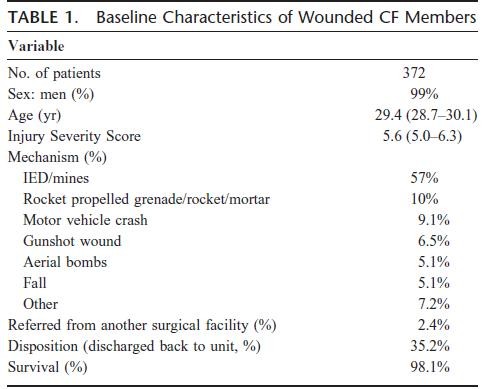
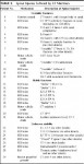
Results: Three hundred seventy-two CF soldiers were injured during the study period and met study criteria. Twenty-nine (8%) had spinal  fractures identified. Of these, 41% (n = 12) were unstable, 31% (n = 9) stable, and 28% indeterminate. Most patients were injured by IEDs (n = 212, 57%). Patients injured by IEDs were more likely to have spinal injuries than those injured by non–IED-related mechanisms (10.4% vs. 2.3%; p < 0.01). IED victims were even more likely to have spinal injuries than patients suffering blunt trauma (10.4% vs. 6.7%; p = 0.02). Prehospital providers were less likely to immobilize the spine in IED victims compared with blunt trauma patients (10% [22 of 212] vs. 23.0% [17 of 74]; p < 0.05).

RachisCAn1.JPGRachisCAn2.JPG

Conclusions: IEDs are a common cause of stable and unstable spinal injuries in the Afghanistan conflict. Spinal immobilization is an underutilized intervention in the battlefield care of casualties in the conflict in Afghanistan. This may be a result of tactical limitations; however, current protocols should continue to emphasize the judicious use of immobilization in these patients.

Lidocaïne intraarticulaire et réduction de luxation d'épaule

 Use of intra-articular lidocaine as analgesia in anterior shoulder dislocation: a review and meta-analysis of the literature

Viktor K et all. Can J Rural Med 2009; 14 (4)

Introduction :

Parmi les blessures affectant des articulations majeures, ce sont celles de l’épaule qui amènent le plus de patients à se présenter à l’urgence d’un hôpital de nos jours. Dans la communauté, l’incidence des blessures à l’épaule est de 11,2 cas  par 100000années-personne. On a généralement recours à la sédation-analgésie perthérapeutique pour faciliter la réduction des luxations antérieures de l’épaule. On
note toutefois certains risques de complications, comme la dépression respiratoire, particulièrement dans certaines populations, d’où la suggestion d’utiliser de la lidocaïne intra-articulaire comme méthode analgésique de rechange.

Méthodes :

Nous avons interrogé les bases de données EMBASE (Ovid) et MEDLINE (PubMed) à partir des mots clés «shoulder, dislocation et/ou reduction» (épaule, luxation et/ou réduction) pour la période allant de la date respective de mise en service des deux bases de données jusqu’à octobre 2008.

Résultats :

Selon la littérature actuelle, il semble que la lidocaïne intra-articulaire  procure au moins le même degré de contrôle et de soulagement de la douleur que la  sédation-analgésie perthérapeutique, tout en réduisant nettement la durée des séjours à l’urgence et le coût du traitement. Il y a lieu de croire que la probabilité de complications serait moindre avec la lidocaïne intra-articulaire.

Conclusion :

 Il faudra approfondir la recherche dans ce domaine, mais les médecins peuvent d’ores et déjà envisager la lidocaïne intra-articulaire comme solution de rechange à la sédation-analgésie perthérapeutique pour la réduction des luxations antérieures de l’épaule.

 Autres références: 1  2  3  4  5