Clicky

Ok

En poursuivant votre navigation sur ce site, vous acceptez l'utilisation de cookies. Ces derniers assurent le bon fonctionnement de nos services. En savoir plus.

22/08/2023

Immobilisation pelvienne improvisée: Pas si mal

Mechanical assessment of proprietary and improvised pelvic binders for use in the prehospital environment


Introduction Pelvic fractures often result from high-energy trauma and are associated with a 10% mortality rate and significant morbidity. Pelvic binders are applied in suspected pelvic injury to stabilise fractured bone, decrease bleeding and potentiate tamponade. A binder must hold the pelvis with sufficient force for this effect to be achieved. This study aims to quantify the ability of proprietary and improvised pelvic binders to hold a target tensile force over time.

Methods The ability of three proprietary and three improvised binders to hold a binding force for 2 hours was tested. A uniaxial materials testing machine was used to tension each binder to 150 N and then hold the displacement for 2 hours; the drop in tension over time was recorded for each binder. The ability to hold tension above 130 N after 2 hours was set as the metric of binder performance.

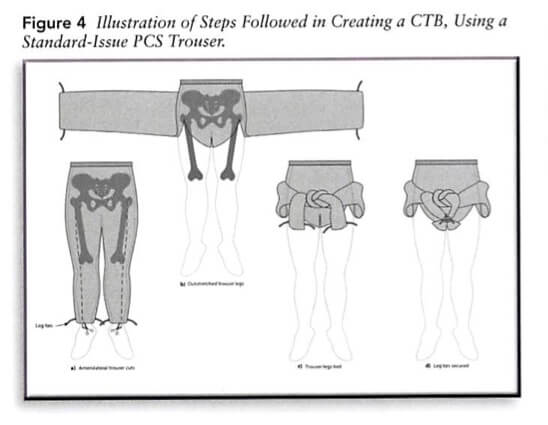
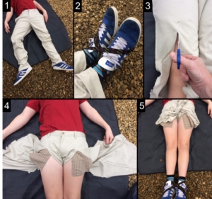
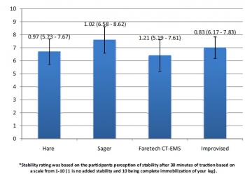
Results The median tension at 2 hours was above 130 N for the SAM Pelvic Sling II and T-POD Pelvic Stabilisation Device and was below 130 N for the Prometheus Pelvic Splint, field-expedient pelvic splint (FES) and the Personal Clothing System-Multi-Terrain Pattern Combat Trousers binders. The tension in the improvised FES after 2 hours was approximately at the target 130 N; however, in 40% of the tests, it held above 130 N.

Conclusions Binders varied in their ability to maintain sufficient tension to treat a pelvic fracture over the 2-hour testing period. The FES performed well under our testing regime; with relatively low cost and weight, it represents a good alternative to proprietary binders for the austere environment.


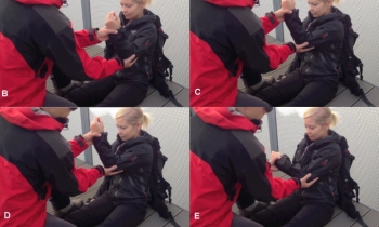
Le Field expedient System: Construit avec un garrot et une attelle SamSplint

6-Figure5-1.png


09/07/2021

Immobilisation pelvienne avec un garrot et une samsplint


bmjmilitary-2021-001838.extract.jpg

08/02/2020

Rachis et extraction de véhicules

An explorative, biomechanical analysis of spine motion during out-of-hospital extrication procedures.

Häske D et Al. Injury. 2020 Feb;51(2):185-192.

------------------------------------

Un travail qui interpelle par ses  implications potentielles sur la manière d'extraire les combattants de véhicules qu'ils soient terrestres ou aéronautiques.

------------------------------------

OBJECTIVES:

The extrication of patients following a road traffic collision is among the basic procedures in emergency medicine. Thus, extrication is a frequently performed procedure by most of the emergency medical services worldwide. The appropriate extrication procedure depends on the patient's current condition and accompanying injuries. A rapid extrication should be performed within a few minutes, and the cervical spine (at least) should be immobilized. To our knowledge, the scientific literature and current guidelines do not offer detailed recommendations on the extrication of injured patients. Thus, the aim of the current study is to compare the effectiveness of spinal stabilization during various out-of-hospital extrication procedures.

METHODS: This is an explorative, biomechanical analysis of spine motion during different extrication procedures on an example patient. Movement of the cervical spine was measured using a wireless human motion tracker. Movement of the thoracic and lumbar spine was quantified with 12 strain gauge sensors, which were positioned paravertebrally on both sites along the thoracic and lumbar spine. To interpret angular movement, a motionscore was developed based on newly defined axioms on the biomechanics of the injured spine.

RESULTS: Self-extrication showed the least spinal movement (overall motionscore sum = 667). Movement in the cervical spine could further be reduced by applying a cervical collar. The extrication by a rescue boa showed comparable results in overall spinal movement compared to the traditional extrication via spineboard (overall motionscore sum = 1862vs. 1743). Especially in the cervical spine, the spinal movement was reduced (motionscore sum = 339 vs. 595). However, the thoracic spine movement was increased (motionscore sum = 812 vs. 432).

Self- extrication without a cervical collar Self- extrication with a cervical collar Rapid extrication Rapid extrication with rescue boa Rapid extrication with a slide board and rescue boa Rapid extrication with a patient transfer sheet
C1 – C7 flexion/extension 25 6 117 132 199 27
rotation 28 6 287 165 216 32
lateral bending 76 19 191 42 65 52
sum 129 31 595 339 480 111
Th1 – Th9 flexion/extension 18 17 69 91 76 24
rotation 157 122 286 598 559 156
lateral bending 65 52 77 123 107 85
sum 240 191 432 812 742 265
Th10 – L2 flexion/extension 21 46 83 109 82 43
rotation 99 185 146 180 114 114
sum 120 231 229 289 196 157
L3 – L5 flexion/extension 38 251 143 254 117 25
rotation 73 93 286 93 178 64
lateral bending 67 67 58 75 117 102
sum 178 411 487 422 412 191
Total sum 667 864 1743 1862 1830 724

CONCLUSION: In case of a suspected cervical spine injury, guided self-extrication seems to be the best option. If the patient is not able to perform self-extrication, using a rescue boa might reduce cervical spinal movement compared to the traditional extrication procedure. Since promising results are shown in the case of extrication using a patient transfer sheet that has already been placed below the driver, future developments should focus on novel vehicle seats that already include an extrication device.

24/07/2019

Examiner un bassin: Peu faible en préhospitalier

Clinical Examination of the Pelvic Ring in the Prehospital Phase.

 
------------------------
Bien que ce travail soit réalisé sur des trauma fermés,  il milite pour un emploi systématiques des ceintures pelviennes dès lors qu'il existe la notion de trauma à haute énergie
------------------------


INTRODUCTION:

Instable pelvic fractures are associated with significant hemorrhage and shock. Instability of the pelvic ring should be tested with the manual compression test (MCT) and instable pelvic ring fractures should prompt mechanical stabilization. However, the accuracy of the prehospital MCT in patients, that sustained a high energetic trauma, is still unknown.

SETTING:

Radboudumc Nijmegen, level 1 trauma center, the Netherlands.

METHODS:

This prospective blind observational study included all patients after a high impact blunt trauma treated by an experienced Helicopter Emergency Medical Service (HEMS) physician. Nominal arranged questionnaires were filled in by the HEMS physician prior to the radiological examination of the patient.

RESULTS:

We included 56 patients of which 11 sustained a pelvic ring fracture. 13 patients were treated with pelvic compression devices, of which only five patients had a pelvic ring fracture. Prehospital performed clinical examination by the HEMS physicians had an overall sensitivity of 0.45 (95% CI 0.16-0.75) and a specificity of 0.93 (95% CI 0.29-0.96).

CONCLUSION:

Pelvic ring instability cannot accurately be diagnosed in the prehospital setting, based on the MCT. The use of the pelvic binder should standard in high impact blunt trauma patients, independently of the MCT or trauma mechanism.

| Tags : pelvis

21/07/2018

Faire avec ce qui est sous la main ?

Improvised first aid techniques for terrorist attacks.


Pelvic-binders-cartoon.jpg

Ceinture pelv improvisée.jpg
 

02/01/2018

Immobilisation cervicale: Pas bon !

Prehospital Spine Immobilization/Spinal Motion Restriction in Penetrating Trauma: a Practice Management Guideline from the Eastern Association for the Surgery of Trauma (EAST)

Trauma Acute Care Surg. 2017 Dec 28


BACKGROUND:

Spine immobilization in trauma has remained an integral part of most emergency medical services (EMS) protocols despite a lack of evidence for efficacy and concern for associated complications, especially in penetrating trauma patients. We reviewed the published evidence on the topic of prehospital spine immobilization or spinal motion restriction in adult patients with penetrating trauma to structure a Practice Management Guideline.

METHODS:

We conducted a Cochrane style systematic review and meta-analysis, and applied GRADE methodology to construct recommendations. Qualitative and quantitative analyses were used to evaluate the literature on the critical outcomes of mortality, neurologic deficit, and potentially reversible neurologic deficit.

RESULTS:

A total of 24 studies met inclusion criteria, with qualitative review conducted for all studies. We used five studies for the quantitative review (meta-analysis). No study showed benefit to spine immobilization with regard to mortality and neurologic injury, even for patients with direct neck injury. Increased mortality was associated with spine immobilization, with RR 2.4 (CI 1.07, 5.41). The rate of neurologic injury or potentially reversible injury was very low, ranging from 0.002 to 0.076 and 0.00034 to 0.055, with no statistically significant difference for neurologic deficit or potentially reversible deficit, RR 4.16 (CI 0.56, 30.89), and RR 1.19 (CI 0.83, 1.70), although the point estimates favored no immobilization.

CONCLUSIONS:

Spine immobilization in penetrating trauma is associated with increased mortality and has not been shown to have a beneficial effect on mitigating neurologic deficits, even potentially reversible neurologic deficits. We recommend that spine immobilization not be used routinely for adult patients with penetrating trauma.

27/09/2016

Ceinture pelvienne improvisée

22/07/2016

De l'usage d'une chèche

Nombre de militaires l'ont adopté car elle protège du soleil, du vent du sable. Tout ceci est connu de longue date notamment des sahariens

 ALG-60-116-R72.jpg

Mais une chèche c'est aussi ET SURTOUT ce qui permet de réaliser une stabilisation pelvienne, un garrot de fortune,un packing de plaie, une immobilisation de fortune, .....

La chèche un équipement important dans le cadre du sauvetage au combat.

Pour vous en convaincre lisez ce document

Et en plus faites une bonne action


14/07/2016

Epaule luxée: Technique de Sool

The effectiveness of a newly developed reduction method of anterior shoulder dislocations; Sool's method

Park MS et Al. Am J Emerg Med. 2016 Apr 9. pii: S0735-6757(16)30036-5. doi: 10.1016/j.ajem.2016.04.01

OBJECTIVE:

Nearly a dozen reduction methods for the treatment of anterior shoulder dislocation have been reported, but the majority are painful and require patients to be in the supine or prone position.

METHODS:

This retrospective cohort study was conducted in a university-affiliated emergency department (ED). Sool's method and traditional shoulder reduction methods (TSRMs) were performed for the patient with anterior shoulder dislocation. Fifty-nine eligible patients were recruited; 35 were treated with TSRMs, wherease 24 were treated with Sool's method.

Luxation Epaule Sool.jpg

clic sur l'image pour voir une vidéo

RESULTS:

The rate of successful reduction was 80% (26/35) in the TSRM group and 75% (18/24) in the Sool's method group (P=.75). The length of stay in the ED was 72.3minutes in the Sool's method group and 98.4minutes in the TSRM group (P=.037). No significant difference was observed between the neurovascular deficit before and after reduction in either group. The procedural time of successfully reduced cases in patients treated by Sool's method was shorter than that of the failed cases (P=.015).

CONCLUSIONS:

Sool's method was as successful as other methods at reducing shoulder dislocation and has demonstrated encouraging results, including significant reduction in length of stay in the ED and unnecessary use of sedation. Sool's method is technically easy and requires only a place to sit and a single operator.

05/04/2016

Immobiliser le rachis: Comme il faut !

 Safety of the lateral trauma position in cervical spine injuries: a cadaver model study

Hyldmo PK et Al. Acta Anaesthesiol Scand. 2016 Mar 7. doi: 10.1111/aas.12714.

__________________________

Bien immobiliser un rachis avec la bonne position. Lire aussi 1, 2, 3

__________________________ 


Background: Endotracheal intubation is not always an option for unconscious trauma patients. Prehospital personnel are then faced with the dilemma of maintaining an adequate airway without risking deleterious movement of a potentially unstable cervical spine. To address these two concerns various alternatives to the classical recovery position have been developed. This study aims to determine the amount of motion induced by the recovery position, two versions of the HAINES (High Arm IN Endangered Spine) position, and the novel lateral trauma position (LTP).
 
Method: We surgically created global cervical instability between the C5 and C6 vertebrae in five fresh cadavers. We measured the rotational and translational (linear) range of motion during the different maneuvers using an electromagnetic tracking device and compared the results using a general linear mixed model (GLMM) for regression.

ImmobCervicale.jpeg

 
Results:
 
In the recovery position, the range of motion for lateral bending was 11.9°. While both HAINES positions caused a similar range of motion, the motion caused by the LTP was 2.6° less (P = 0.037). The linear axial range of motion in the recovery position was 13.0 mm. In comparison, the HAINES 1 and 2 positions showed significantly less motion ( 5.8 and 4.6 mm, respectively), while the LTP did not (4.0 mm, P = 0.067).
 
Conclusion: Our results indicate that in unconscious trauma patients, the LTP or one of the two HAINES techniques is preferable to the standard recovery position in cases of an unstable cervical spine injury.

| Tags : rachis

Victimes d'explosion: Pas de collier cervical

Cervical spine injuries in civilian victims of explosions: Should cervical collars be used?

Klein Y et Al. J Trauma Acute Care Surg. 2016 Mar 18. [Epub ahead of print] 

---------------------------------------------------

En contexte de terrorisme civil, la pose d'un collier cervical apparaît plutôt inutile et source de perte de temps.

---------------------------------------------------

BACKGROUND:
Semi-rigid cervical collars (SRCC) are routinely applied to victims of explosions as part of the pre-hospital trauma protocols. Previous studies have shown that the use of SRCC in penetrating injuries is not justified due to the scarcity of unstable cervical spine injuries and the risk of obscuring other neck injuries. Explosion can inflict injuries by fragments penetration, blast injury, blunt force and burns. The study purpose was to determine the occurrence of cervical spine instability without irreversible neurological deficit and other potentially life threatening non-skeletal neck injuries among victims of explosions. The potential benefits and risks of SRCC application in explosion related injuries (ERI) was Evaluated.

METHODS:
a retrospective cohort study of all explosions civilian victims admitted to Israeli hospitals during the years 1998-2010. Data collection was based on the Israeli national trauma registry and the hospital records, and included demographic, clinical and radiological details of all patients with documented cervical spine injuries.

RESULTS:
the cohort included 2,267 patients. All of them were secondary to terrorist attacks. SRCC was applied to all the patients at the scene. Nineteen patients (0.83%) had cervical spine fractures. Nine patients (0.088%) had unstable cervical spine injury. All but one had irreversible neurological deficit on admission. 151 patients (6.6%) had potentially life threatening penetrating non-skeletal neck injuries.

CONCLUSIONS:
Unstable cervical spine injuries secondary to explosion are extremely rare. The majority of unstable cervical spine fractures were secondary to penetrating injuries, with irreversible neurological deficits on admission. The application of SRCC did not seem to be of any benefit in these patients and might pose a risk of obscuring other neck injuries. We recommend that SRCC will not be used in the pre-hospital management of victims of explosions.

LEVEL OF EVIDENCE:
Retrospective observational study, level III.

| Tags : immobilisation

08/03/2016

Epaule luxée ! Réduire seul

Simple self-reduction method for anterior shoulder dislocation

Reiner Wirbel et al. Journal of Acute Disease (2014)207-210


C'est la plus fréquente des luxations. La diversité des méthodes de réduction illustre bien la difficulté qu'il peut y avoir à la réduire sans avoir recours à une anesthésie/sédation. C'est encore plus vrai en cas d'isolement extrême. Une méthode à connaître. 

Reduc LAI.jpg

ReduLAI2.jpg

18/02/2016

Stabilisation pelvienne: Les genoux sont importants

Pelvic pressure changes after a fracture: A pilot cadaveric study assessing the effect of pelvic binders and limb bandaging

Morris R et Al. Injury  http://dx.doi.org/10.1016/j.injury.2015.12.009

-------------------------

Stabiliser un bassin pour lequel il existe une suspicion de fracture est une des composantes majeures du sauvetage au combat. Ceci vise à réduire la mortalité par hémorragie non garrotable ou non comprimable. Il existe des ceintures spécifiques pour cela. Ce travail est intéressant car il illustre la complémentarité des ceintures pelviennes ET de l'immobilisation des genoux. Cette dernière permet à elle seule d'augmenter la pression intra-abdominale et partant probablement de réduire le saignement. Donc n'oublier pas d'immobiliser les genoux. Que vous disposez ou pas de ceinture pelvienne, cela sert.

-------------------------

Objectives: Pelvic binders are a life-saving intervention for hypovolaemic shock following displaced pelvic fractures, thought to act through increasing intra-pelvic pressure to reduce venous bleeding. This cadaveric study assesses changes in intra-pelvic pressure with different binders augmented by bandaging the thighs to recruit the femora as levers to close the pelvis. Access to femoral vessels via an in situ binder was also assessed.

Methods: Two embalmed cadavers were used with unstable pelvic injuries (OA/OTA 61-C1) created through disrupting the pelvic ring anteriorly and posteriorly. To measure intravesical pressure, which reflects intra-pelvic pressure, a supra-pubic catheter was inserted and connected to a water manometer whilst a spigot was placed in a urethral catheter to reduce leakage of fluid. The common and superficial femoral arteries were dissected in the left groin for each specimen prior to any intervention to allow inspection following binder application. A SAM pelvic sling II, Trauma Pelvic Orthotic Device (T-POD), Prometheus pelvic splint and an improvised pelvic binder were used on each cadaver, with each applied following lower limb bandaging with the knees slightly flexed. The groins were then inspected to assess if the femoral vessels were visible. Statistical analysis was performed in SPSS using a paired samples t test to determine if any difference existed between initial pelvic pressure in specimens compared to pressures with bandaging on and binders applied.

Results: Bandaging the lower limbs alone produced a significant increase in both peak and steady mean intra-pelvic pressure, 15.69 cmH2O and 12.38 cmH2O, respectively, compared to the baseline pressure, 8.73 cmH2O (p = 0.002 and p = 0.001, respectively). Applying the pelvic binder with the bandaging in place increased intra-pelvic pressure compared to the baseline (peak pressure of 25.38 cmH2O (p < 0.001) and steady pressure of 15.13 cmH2O (p = 0.003)). Steady mean pressures between bandaging alone and bandaging with the binder applied were not significantly different (p = 0.09), whilst the peak pressures were significantly greater when the binder was applied (p = 0.005). The improvised binder and T-POD both required cutting to access the femoral vessels which resulted in decreasing efficacy.

Conclusions: Intra-pelvic pressure was significantly increased through bandaging the lower limbs alone, and this represents a simple measure to increase intra-pelvic pressure and therefore efficacy of the binder. Access to the femoral vessels varied with binder type and represents an important consideration in polytrauma patients.

19/12/2015

Amputé des jambes: Le bassin aussi !

The incidence of pelvic fractures with traumatic lower limb amputation in modern warfare due to improvised explosive devices

Cross AM et Al. J R Nav Med Serv 2014;100(2):152-6

---------------------------------------------

Excepté l'extraction d'urgence de blessés sous le feu, la prise en charge den cas d'amputation traumatique doit inclure la forte probabilité de traumatisme du bassin. Une utilisation large des immobilisations pelviennes doit donc être à l'esprit. On rappelle simplement la gravité et la difficulté de prise en charge des hémorragies liées aux fractures de bassin.

---------------------------------------------

AIMS:

A frequently-seen injury pattern in current military experience is traumatic lower limb amputation as a result of improvised explosive devices (IEDs). This injury can coexist with fractures involving the pelvic ring. This study aims to assess the frequency of concomitant pelvic fracture in IED-related lower limb amputation.

METHODS:

A retrospective analysis of the trauma charts, medical notes, and digital imaging was undertaken for all patients arriving at the Emergency Department at the UK military field hospital in Camp Bastion, Afghanistan, with a traumatic lower limb amputation in the six months between September 2009 and April 2010, in order to determine the incidence of associated pelvic ring fractures.

RESULTS:

Of 77 consecutive patients with traumatic lower limb amputations, 17 (22%) had an associated pelvic fracture (eleven with displaced pelvic ring fractures, five undisplaced fractures and one acetabular fracture). Unilateral amputees (n = 31) had a 10% incidence of associated pelvic fracture, whilst 30 % of bilateral amputees (n = 46) had a concurrent pelvic fracture. However, in bilateral, trans-femoral amputations (n = 28) the incidence of pelvic fracture was 39%.

PelvicFractures.jpg

BKA - Below knee amputation; AKA - Above knee amputation

CONCLUSIONS:

The study demonstrates a high incidence of pelvic fractures in patients with traumatic lower limb amputations, supporting the routine pre-hospital application of pelvic binders in this patient group

PelvicImmobilisation.jpg

04/08/2015

Collier cervical: Pas utile ?

Cervical spine injury in dismounted improvised explosive device trauma

Taddeo j et Al. Can J Surg. 2015 Jun; 58(3 Suppl 3): S104–S107

-------------------------------

La mise en place d'un collier cervical est une mesure classique en traumaotlogie routière. Elle n'est cependant pas recommandée en cas de traumatisme d'origine ballistique. La probabilité de lésions instables du rachis semble faible surtout si il s'agit de blessures survenues lors de combat à pied et sans atteinte crânienne associée. L'étude présentée conforte cette position.

-------------------------------

Background

The injury pattern from improvised explosive device (IED) trauma is different if the target is in a vehicle (mounted) or on foot (dismounted). Combat and civilian first response protocols require the placement of a cervical collar on all victims of a blast injury.

Methods

We searched the Joint Theatre Trauma Registry (JTTR) and the Role 3 Hospital, Kandahar Airfield (KAF) database from Mar. 1, 2008, to May 31, 2011. We collected data on cervical fracture; head injury; traumatic amputation; initial blood pressure, pulse, injury severity score (ISS), Glasgow Coma Scale (GCS) score and base excess; and patient demographic information.

Results

The concordance rate between JTTR and KAF databases was 98%. Of the 15 693 admissions in JTTR, 326 patients with dismounted IED injuries were located. The rate of cervical collar prehospital placement was 7.6%. Cervical fractures were found in 19 (5.8%) dismounted IED victims, but only 4 (1.2%) were considered radiographically unstable. None of these 19 patients had prehospital placement of a collar. Patients with cervical spine fractures were more severely injured than those without (ISS 18.2 v. 13.4; GCS 10.1 v. 12.5). Patients with head injuries had significantly higher risk of cervical spine injury than those with no head injury recorded (13.6% v. 3.9%). No differences in frequency of cervical spine injury were found between patients who had associated traumatic amputations and those who did not (5.4% v. 6.0%).

Conclusion

Dismounted IED is a mechanism of injury associated with a low risk for cervical spine trauma. A selective protocol for cervical collar placement on victims of dismounted IED blasts is possible and may be more amenable to combat situations.

| Tags : rachis

19/07/2015

Immobilisation: Matériel spécifique ?

Improvised traction splints: a wilderness medicine tool or hindrance?

Weichenthal L et Al. Wilderness Environ Med. 2012 Mar;23(1):61-4

------------------------------------------------

Immobiliser un membre le plus tôt possible et ce de manière efficace est un des enjeux de la mise en condition d'évacuation. Il s'agit là de prévenir le mieux possible toute complication secondaire, de réduire le saignement et la douleur. Pour cela existe de nombreux équipements proposés par l'industrie. Miass sont ils vraiment utiles ? Pas si évident si l'on en croit le document proposé. Il pourrait exister à l'évidence des moyens simples et efficaces surtout moins coûteux d'atteindre ces objectifs.

------------------------------------------------

OBJECTIVES:

To investigate whether a traction splint made from improvised materials is as efficacious as commercially available devices in terms of traction provided and perceived comfort and stability.

METHODS:

This was a prospective randomized crossover study utilizing 10 healthy, uninjured volunteers. The subjects were randomized to be placed in 4 different traction devices, in differing order, each for 30 minutes. Three of the traction splints are commercially available: The HARE, Sager, and Faretech CT-EMS. The fourth traction device was an improvised splint made as described in Medicine for the Backcountry: A Practical Guide to Wilderness First Aid. At the end of 30 minutes the pounds of force created by each device was measured. The volunteers were also asked at that time to subjectively report the comfort and stability of the splint separately on a scale from 1 to 10.

RESULTS:

All traction splints performed similarly with regard to the primary outcome measure of mean pounds of traction created at the end of 30 minutes of application with results ranging from 10.4 to 13.3 pounds. There was little difference reported by participants in regard to stability or comfort between the 4 traction devices.

ImproSplint1.jpg

ImproSplint2.jpg

CONCLUSIONS:

In this small pilot study, an improvised traction splint was not inferior to commercially available devices. Further research in needed in this area.

| Tags : immobilisation

07/07/2015

Quelle attelle de fémur ?

Evaluation of commercially available traction splints for battlefield use

Studer NM et All. J Spec Oper Med. 2014 Summer;14(2):46-55

------------------------------------------------------

L'immobilisation d'une fracture de fémur nécessite idéalement une traction. Les attelles de type thomas-lardenois ne sont pas utilisables en préhospitalier. La classique attelle de Donway n'est pas idéale dès lors que l'on est en combat à pied. Bien que l'intérêt de telles attelles reste discuté (1) , nous disposons à la nomenclature  de l'atelle Faretec CT6. Le document présenté vante de manière très surprenante les qualités de l'attelle de slishman. Certes cette dernière semble plus rapide à poser mais avec un taux d'échec beaucoup plus important. La CT6 reste donc un excellent choix, c'est d'ailleurs ce qui est également dit dans ce document qui parait quelque peu orienté dans ces conclusions. 

------------------------------------------------------

Background: Femoral fracture is a common battlefield injury with grave complications if not properly treated. Traction splinting has been proved to decrease morbidity and mortality in battlefield femur fractures. However, little standardization of equipment and training exists within the United States Armed Forces. Currently, four traction splints that have been awarded NATO Stock Numbers are in use: the CT-6 Leg Splint, the Kendrick Traction Device (KTD), the REEL Splint (RS), and the Slishman Traction Splint (STS).

Objective: The purpose of this study was to determine the differences between the four commercially available traction devices sold to the U.S. Government.

Methods: After standardized instruction, subjects were timed and evaluated in the application of each of the four listed splints. Participant confidence and preferences were assessed by using Likertscaled surveys. Free response remarks were collected before and after timed application.

Results: Subjects had significantly different application times on the four devices tested (analysis of variance [ANOVA], p < .01). Application time for the STS was faster than that for both the CT-6 (t-test, p < .0028) and the RS (p < .0001). Subjects also rated the STS highest in all post-testing subjective survey categories and reported significantly higher confidence that the STS would best treat a femoral fracture (p < .00229).

Conclusions: The STS had the best objective performance during testing and the highest subjective evaluation by participants. Along with its ability to be used in the setting of associated lower extremity amputation or trauma, this splint is the most suitable for battlefield use of the three devices tested

| Tags : immobilisation

19/05/2015

Luxation d'épaule: Une méthode simple ?

Reduction of Acute Shoulder Dislocations in a Remote Environment: A Prospective Multicenter Observational Study

Bokor-Billmann T. et Al. Wilderness Environ Med. 2015 Mar 27. pii: S1080-6032(15)00006


OBJECTIVE:

Acute dislocations of the glenohumeral joint are common in wilderness activities. Emergent reduction should take place at the site of trauma to reduce the patient's pain and the risk of vascular and neurological complications. A limited number of reduction methods are applicable in remote areas. The aim of this study is to present our method of reduction of anterior shoulder luxation that is easily applicable in remote areas without medication, adjuncts, and assistants and is well tolerated by patients.

METHODS:

A prospective observational study was conducted during a 5-year period. The patients included underwent closed manual reduction with our technique. After each reduction, the physician who performed the reduction completed a standardized detailed history, and reexamined the patient (for acute complications). The patients were contacted 6 months after the trauma to investigate long-term postreduction complications.

Diagram depicting the steps of our reduction procedure. The practitioner holds the patient’s wrist with the left hand (in the case of a left shoulder dislocation) and the patient’s elbow with the right hand.

Réduction LAI Epaule.jpg


(B) With the elbow in 90° of flexion, the glenohumeral joint is flexed forward to 90°. (C) While still in flexion, the glenohumeral joint is adducted until the elbow reaches the midline of the body; it is important to continue this movement until this landmark is completely reached. (D) Then, internal rotation of the shoulder is performed. During this step, the patient’s elbow must stay at the landmark described above. At 25° to 30° of rotation, a mild resistance is usually encountered. (E) The last step of the maneuver consists of applying a constant internal rotation pressure to overcome this mild resistance without pain. Reduction is usually achieved at approximately 30° of internal rotation.

RESULTS:

Reduction was achieved with our method in 39 (100.0%) of 39 patients. The mean pain felt during our reduction procedure was rated 1.7 ± 1.4 (on a scale of 10) using the visual analog scale scoring system. No complications were noted before or after the reduction attempts. We did not find any long-term complications.

CONCLUSIONS:

The reduction method presented in the present study is an effective method for the reduction of acute shoulder luxations in remote places. Our data suggest that this method could be applied for safe and effective reduction of shoulder dislocation.


| Tags : réduction

26/03/2015

Techniques d'immobilisations

ImmobUrgences.jpg


clic sur l'image pour accéder aux documents

| Tags : immobilisation

03/03/2015

Stabilisation du rachis: C'est particulier en milieu hostile

Wilderness medical society: Practice Guidelines for spine immobilization in the austere environment.
Quinn R. et Al. Wilderness Environ Med. 2013 Sep;24(3):241-52

Recommandation 1 (grade:1C) :

Neutral alignment should be restored and maintained with light or moderate manual cervical traction during extrication, unless such a maneuver is met with resistance, increased pain, or new or worsening neurologic deficit.

Recommandation 2 (grade:1C):

Patients requiring extrication, when the cervical spine cannot be cleared before extrication, should be placed in a cervical collar and allowed to exit the situation under their own volition if alert and reliable. Otherwise extrication should be performed with a KED (or similar device) plusc ervical collar,and the immobilized patient moved in a sitting position onto a long spine board, vacuum mattress, orsimilar device.

Recommandation 3 (grade:1C):

The lift and slide transfer with trap squeeze is preferred to the log roll when transferring patients to and from a backboard

Recommandation 4 (grade:2C):

Spinal immobilization should be considered in patients with evidence of spinal injury, including those with neurologic injury, and those patients who have experienced severe trauma and are unconscious or exhibit altered mental status.

Recommandation 5 (grade:2B):

The cervical collar (or improvised equivalent) should beconsidered one ofs everal tools available to aid in immobilization of the cervical spine. It should not be considered adequate immobilization in and of itself, nor should it beconsidered necessary if adequate immobilization can be accomplished by other means, or if the presence of the collar in itself compromises emergent patient care.

Recommandation 6 (grade:1B):

Use of the cervical collar is contraindicated in ankylosing spondylitis. Patients with suspected injury should hav etheir neck supported in a position of comfort 

Recommandation 6 (grade:1C):

Vacuum mattress provides superior immobilization, with or without a standard cervical collar, and improved patient comfort (with corresponding decreased risk of pressure sores) and is preferred over a backboard for immobilization of either the entire spine or specific segments of concern.

 Recommandation 6 (grade:1A):

 Appropriately trained personnel, using either the NEXUS criteria or the Canadian C- spine rule, can safely and effectively make decisions in the prehospital setting about whether or not ac ervical spine should be immobilized.

Recommandation 7(grade:1B):

Spinal immobilization should not be performed in the presence of penetrating trauma


spine.jpeg.jpg 

Clic sur l'image pour accéder au document