Clicky

Ok

En poursuivant votre navigation sur ce site, vous acceptez l'utilisation de cookies. Ces derniers assurent le bon fonctionnement de nos services. En savoir plus.

29/06/2013

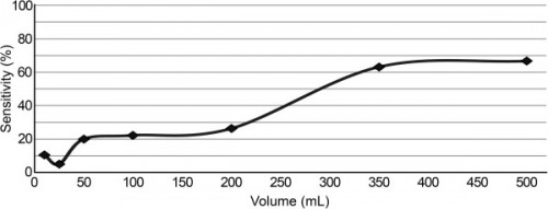
Pneumothorax: A partir de quel volume d'air le dépiste-t-on ?

The intrapleural volume threshold for ultrasound detection of pneumothoraces: An experimental study on porcine models

Oveland NP et All. Scand J Trauma Resusc Emerg Med. 2013; 21: 11.

Pneumo.jpg

Tous les pneumothorax sont dépistés à l'échographie pour des volumes d'air de moins de 50 ml, de manière bien plus précoce que l'analyse d'une radiographie comme le montre la figure ci-dessus

| Tags : pneumothorax

24/06/2013

Pneumothorax: Echographie

 Husain LF et All. Sonographic diagnosis of pneumothorax.

J Emerg Trauma Shock 2012;5:76-81

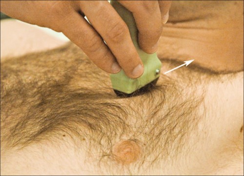
Position de la sonde 

Pneumothorax_Sonde.jpg

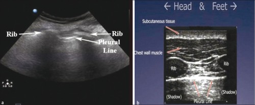
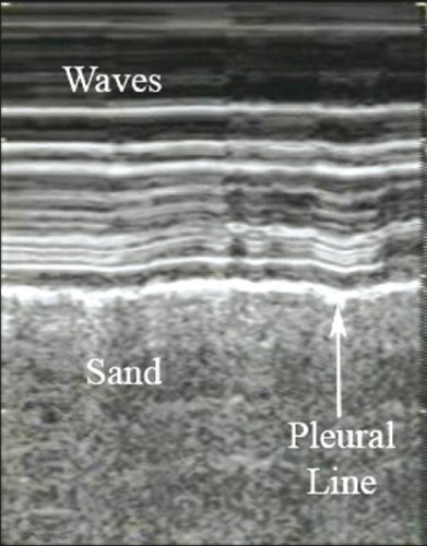
Images normales

Le glissement pleural        Vidéo

La chauve-souris

Pneumothorax_Images Noramales1.jpg

Le bord de plage

Pneumothorax_Images Normales2.jpg

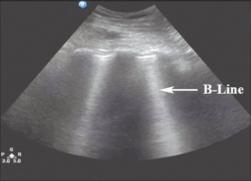
Les lignes B

Pneumothorax_Images Normales3.jpg


Images Anormales attestant du pneumothorax

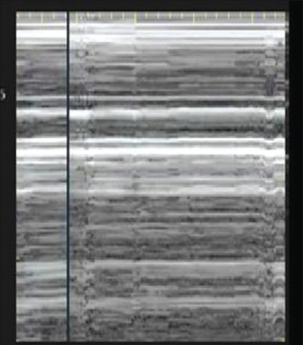
Pas de glissement                                                        Vidéo 

Le signe du Barcode

Pneumothorax_Images PasNormales1.jpg

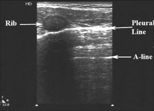
Les lignes A

Pneumothorax_Images PasNormales2.jpg

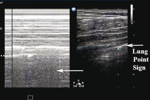
Le point poumon

Pneumothorax_Images PasNormales3.jpg

Le pouls pulmonaire

lung.left-lung-pulse.M-mode-300x225.jpg


Pour aller plus loin:

Un cours commenté

Un diaporama du BMP


20/04/2013

Pneumothorax compressif: Comprendre


01/10/2012

Epaisseur de paroi thoracique: Le point chez le militaire français

Mesure de l’épaisseur de la paroi thoracique chez des militaires français :

Quelle technique pour l’exsufflation du pneumothorax compressif lors du sauvetage au combat?

A propos d’une étude descriptive réalisée du 1er Mars au 15 Avril 2010

au 7ème Bataillon de Chasseurs Alpins de Bourg Saint Maurice.

LAMBLIN A. Thèse Lyon 2012


Le diaporama de présentation      :       EpThoDiaporama.pdf

La thèse                                          :       Lien

20/09/2012

Pneumothorax: La sonde d'écho à quel endroit ?

 Anatomical distribution of traumatic pneumothoraces on chest computed tomography: implicationsfor ultrasound screening in the ED

Mennicke M et alll. Am J Emerg Med 2012 Sep;30(7):1025-31

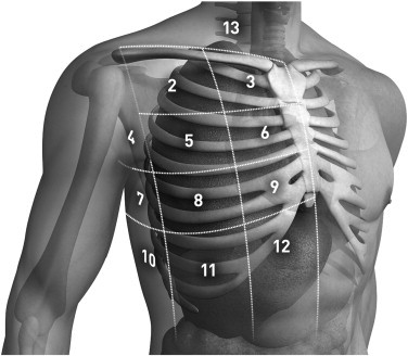
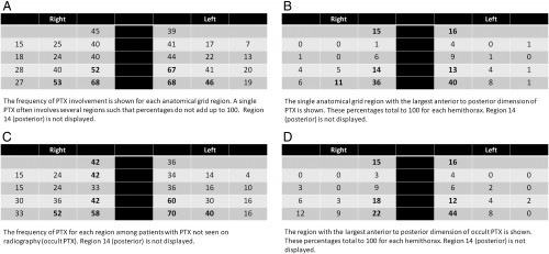
Un travail qui précise les zones optimales d'exploration de la région thoracique pour l'identiifcation optimale des pneumothorax traumatiques.

----------------------------------------------------------------------------------------

OBJECTIVES:

We sought to assess the anatomical distribution of traumatic pneumothoraces (PTXs) on chest computed tomography (CT) to develop an optimized protocol for PTX screening with ultrasound in the emergency department (ED).

METHODS:

We performed a retrospective review of all chest CTs performed in one ED between January 2005 and December 2008 according to presence, location, and size of PTX. Pneumothoraces were then measured and categorized into 14 anatomical regions for each hemithorax.


RESULTS:

A total of 277 (3.8%) PTXs were identified, with 26 bilateral PTX, on 3636 chest CTs performed during the study period. Etiology was blunt (85%) or penetrating trauma (15%). Eighty-three (45%) PTXs were radiographically occult on initial chest x-ray. One hundred eighty-three (66%) PTX had no chest tube at the time of CT. For both hemithoraces, the distribution demonstrated increasing PTX frequency and size from lateral to medial and from superior to inferior. Region 12 (parasternal, intercostal spaces [ICS] 7-8) was involved in 68% of PTX on either side; region 9 (parasternal, ICS 5-6), in 67% on the left and in 52% on the right; and region 11 (lateral to midclavicular line, ICS 7-8), in 46% on the left and in 53% on the right. The largest anterior-to-posterior PTX dimension was seen in region 12.

zones thorax.jpg

Pneumothorax.jpg

CONCLUSIONS:

Our results indicate that 80.4% of right- and 83.7% of left-sided traumatic PTXs would be identified by scanning regions 9, 11, and 12. These findings suggest that a standardized protocol for PTX screening with ultrasound should include these regions.

----------------------------------------------------------------------------------------


23/02/2012

Exsufflation à l'aiguille: Le risque est rare mais réel !

Subclavian artery laceration: A serious complication of needle decompression

Riwoe D et all. Emergency Medicine Australasia (2011) 23, 651–653

Un article qui rapporte la survenue d'une dialcération de l'artère sous clavière après exsufflation à l'aiguille par voie antérieure. Les auteurs militent pour la réalisation d'une thoracostomie au doigt par voie latérale ou après repérage échographique sinon l'emploi de repère cutanés préconisés par Wax et coll. qui font appel à la reconnaissance de la fourchette sternale et la ligne médio-thoracique mamelonnaire.

06/01/2012

Passer par le côté, c'est mieux

J Trauma. 2011 Nov;71(5):1099-103; discussion 1103.

Optimal positioning for emergent needle thoracostomy: a cadaver-based study.

Source

Division of Trauma and Surgical Critical Care, University of Southern California, Los Angeles, California 90033-4525, USA. kinaba@surgery.usc.edu

Abstract

BACKGROUND:

Needle thoracostomy is an emergent procedure designed to relieve tension pneumothorax. High failure rates because of the needle not penetrating into the thoracic cavity have been reported. Advanced Trauma Life Support guidelines recommend placement in the second intercostal space, midclavicular line using a 5-cm needle. The purpose of this study was to evaluate placement in the fifth intercostal space, midaxillary line, where tube thoracostomy is routinely performed. We hypothesized that this would result in a higher successful placement rate.

METHODS:

Twenty randomly selected unpreserved adult cadavers were evaluated. A standard 14-gauge 5-cm needle was placed in both the fifth intercostal space at the midaxillary line and the traditional second intercostal space at the midclavicular line in both the right and left chest walls. The needles were secured and thoracotomy was then performed to assess penetration into the pleural cavity. The right and left sides were analyzed separately acting as their own controls for a total of 80 needles inserted into 20 cadavers. The thickness of the chest wall at the site of penetration was then measured for each entry position.

RESULTS:

A total of 14 male and 6 female cadavers were studied. Overall, 100% (40 of 40) of needles placed in the fifth intercostal space and 57.5% (23 of 40) of the needles placed in the second intercostal space entered the chest cavity (p < 0.001); right chest: 100% versus 60.0% (p = 0.003) and left chest: 100% versus 55.0% (p = 0.001). Overall, the thickness of the chest wall was 3.5 cm ± 0.9 cm at the fifth intercostal space and 4.5 cm ± 1.1 cm at the second intercostal space (p < 0.001). Both right and left chest wall thicknesses were similar (right, 3.6 cm ± 1.0 cm vs. 4.5 cm ± 1.1 cm, p = 0.007; left, 3.5 ± 0.9 cm vs. 4.4 cm ± 1.1 cm, p = 0.008).

CONCLUSIONS:

In a cadaveric model, needle thoracostomy was successfully placed in 100% of attempts at the fifth intercostal space but in only 58% at the traditional second intercostal position. On average, the chest wall was 1 cm thinner at this position and may improve successful needle placement. Live patient validation of these results is warranted.

26/11/2011

Exsufflation: 5 cm suffisent VRAIMENT !

Determination of the appropriate catheter length for needle thoracostomy by using computed tomography scans of trauma patients in Japan
Injury, In Press, Corrected Proof, Available online 24 December 2010
Takeshi Yamagiwa, Seiji Morita, Rie Yamamoto, Tomoko Seki, Katsuhiko Sugimoto, Sadaki Inokuchi
PDF (164 K) 

Background

Previous studies reported a high failure rate in relieving tension pneumothorax by needle thoracostomy, because the catheter was not sufficiently long to access the pleural space. The Advanced Trauma Life Support guideline recommends needle thoracostomy at the second intercostal space in the middle clavicular line using a 5.0-cm catheter, whereas the corresponding guideline in Japan does not mention a catheter length. It is necessary to measure the chest wall thickness (CWT) and determine the appropriate catheter length taking the differences of habitus in race and region into consideration. This study was designed to analyse CWT in Japanese trauma patients by computed tomography and to determine the percentage of patients whose pleural space would be accessible using a 5.0-cm catheter.

Patients and methods

We performed a retrospective review of chest computed tomography of 256 adult Japanese trauma patients who were admitted to the level 1 trauma centre of Tokai University Hospital in Kanagawa, Japan between January and July 2008. In 256 patients, the CWT at 512 sites (left and right sides) was measured by chest computed tomography at the second intercostal space in the middle clavicular line. The frequency of measurement sites <5.0 cm was calculated simultaneously.

The samples were divided according to gender, side (left and right), abbreviated injury scale (<3, ≧3), arm position during examination (up/down), and the existence or non-existence of associated injuries (pneumothorax, subcutaneous emphysema, and fracture of the sternum and ribs); the CWT of each group was compared.

Results

The mean CWT measured in 192 males and 64 females was 3.06 ± 1.02 cm. The CWT values at 483 sites (94.3%) were less than 5.0 cm. The CWT of females was significantly greater than that of males (3.66 cm vs. 2.85 cm, p < 0.0001), and patients with subcutaneous emphysema had greater CWTs than those without it (4.16 cm vs. 3.01 cm, p < 0.0001).

Conclusion

The mean CWT at the second intercostal space in the middle clavicular line was 3.06 cm. It is likely that over 94% of Japanese trauma patients could be treated with a 5.0-cm catheter.

26/12/2010

Drainage thoracique en images

Le drainage thoracique en condition de combat doit rester l'exception. Il peut se justifier en cas d'échec ou d'insuffisance de l'exsufflation ou de la thoracostomie au doigt. Il peut également s'envisager selon les conditions tactiques si le délai d'EVASAN es long. Le document ci-après présente de manière très didactique la réalisation de ce geste pas si anodin qu'il n'y paraît.

http://www.copacamu.org/IMG/pdf/3-roch.pdf